Summary Anatomy and Fysiology
Anatomie en fysiologie (AB_1176)
Vrije Universiteit Amsterdam
Aanbevolen voor jou


Reacties
Preview tekst
Summary Anatomy and Physiology (AB_1176)
Fundamentals of Anatomy and Physiology; Martini
H20 The Heart
20: The heart is a four chambered organ that pumps blood through the systemic and pulmonary circuits Heart: hollow muscular organ in thoracic cavity – pulmonary (lung, short): carries blood to and from the gas exchange surfaces of the lungs— and systemic circuit (long, to organs): transports blood to and from the rest of the body - Arteries carry blood away from the heart, veins return blood—great vessels are connected to the heart; capillaries (smallest arteries) are interconnected; their thin wall permits the exchange of nutrients, dissolved gasses, and waste. - Four muscular chambers: (1) right atrium receives blood from systemic circuit and passes it to (2) right ventricle pumps blood into pulmonary circuit (3) left atrium collects blood hereof and empties into (4) left ventricle pumps blood into systemic circuit >> first atria contract, then ventricles (equal volumes) - Great vessels connected to superior end of the heart at its base; third costal cartilage—inferior tip of the heart is the apex >> heart sits in anterior portion of the mediastinum, between two pleural cavities >> mediastinum contains great vessels, thymus, esophagus, and trachea
- Walls of heart: endocardium (inner layer), myocardium (spiral bundle of cardiac muscle cells), pericardium (fibrous and serous membrane) is two layered: parietal and visceral layer which are separated by fluid-filled pericardial cavity 1. Pericardium >> visceral layer of serous pericardium (epicardium) covers surface of the heart: exposed mesothelium and underlying layer of areolar connective tissue >> parietal layer of serous pericardium: outer dense fibrous layer, areolar layer, and an inner mesothelium 2. Myocardium forms atria and ventricles – contains cardiac muscle cells, connective tissue, blood vessels, and nerves—muscle bundles encircle the great vessels – deeper muscle layers spiral around 3. Endocardium covers inner surfaces of the heart and heart valves – simple squamous epithelium and underlying areolar tissue
- Cardiac skeleton is supporting (crisscrossing) limits the spread of action potentials: four dense bands of tough elastic tissue that stabilize positions of heart valves and ventricular muscle
- Pericardium surrounds the heart; pathogens can infect => inflammation => pericarditis (surfaces rub against one another, making a scratching sound ==> commonly results in increased production of pericardial fluid -> collects in cavity; restricting movement of the heart => cardiac tamponade (also from traumatic injuries) > outer fibrous pericardium; dense network of collagen fibers that stabilize position > inner serous pericardium; outer parietal and inner visceral (epicardium) – potential, fluid-filled space: pericardial cavity – pericardial fluid secreted by pericardial membranes acts as lubricant -> reducing friction
Two atria have relatively thin muscular walls and are highly expandable – outer portion deflates => lumpy, wrinkled flap: auricle - Coronary sulcus: deep groove, marks border between atria and ventricles: anterior interventricular sulcus and posterior interventricular sulcus are shallower => boundary between left and right ventricles – fat in sulci - Connective tissue of the heart includes collagen and elastic fibers (sheath) – fibrous cross-links/ struts interwoven into sheets (1) provide physical support of for myocardium (2) help distribute forces of contraction (3) add strength and prevent overexpansion of the heart (4) provide elasticity -> original shape and size
- Chambers are separated by septa – atria are separated by (thicker) interventricular septum – valves: covered openings >> two atrioventricular (AV) valves: folds of fibrous tissue; extend into openings between (and from: one direction) atria and ventricles >> two semilunar valves between ventricles and their great vessels
- Right atrium receives blood from superior vena cava (from head, neck, upper limbs, and chest) and inferior vena cava (from rest of trunk, viscera, and lower limbs) – no valves between venae cavae and right atrium
Formane ovale penetrates interatrial septum and connects two atria of the fetal heart => permits blood flow from right atrium to left atrium while lungs are developing => closes at birth => sealed >> fossa ovalis remains at this site in the adult heart - Posterior walls of right atrium and interatrial septum have smooth surfaces – muscular ridges: pectinate muscles >> blood travels through flaps: cusps; part of tricuspid valve (right atrioventricular (AV) valve) – free edge of each cusp attached to chordae tendineae that originate at the papillary muscles - Internal surface of right ventricle contains series of muscular ridges: trabeculae carneae – moderator band delivers stimulus for contraction to the papillary muscles => begin tensing the chordae tendineae before the rest of the ventricle contracts – conus arteriosus ends at pulmonary arteriosus; consists of three semilunar cusps (thick connective tissue) - Blood flowing from right ventricle passes through this valve into pulmonary trunk => into left pulmonary arteries and right pulmonary arteries – vessels branch within lungs before supplying the capillaries
Respiratory capillaries unite => four pulmonary veins – left atrium receives blood from two left and two right pulmonary veins – mitral valve guards the entrance to the left ventricle; contains two cusps - Left ventricle had no moderator bands – pair of large papillary muscles tenses the chordae tenidinae that anchor the cusps of the mitral valve and prevent blood from flowing back into left atrium >> through aortic valve (adjacent each side: saclike expansions: aortic sinuses)-> enters ascending aorta -> through aortic arch and into descending aorta – pulmonary trunk attached to aortic arch by ligamentum arteriosum (= fibrous band) - Right and left ventricles have significant structural differences – left ventricle much larger and has walls thicker => enables left ventricle to push blood through body’s extensive systemic circuits (4-6x more pressure) >> lungs are close to heart; pulmonary blood vessels are relatively short and wide; no hard pushing => right ventricle resembled pouch attached to the massive wall of left ventricle – moves blood efficiently with minimal effort - Left ventricle contracts: it shortens and narrows => (1) distance between base and apex decreases (2) diameter of ventricular chamber decreases – pressure generated is enough to open aortic valve and eject blood into ascending aorta; contraction; bulges into right ventricular cavity => increases pumping efficiency of right ventricle
Atrioventricular (AV) valves prevent backflow of blood from ventricles to atria when ventricles are contracting – chordae tendineae are loose; no resistance – when it contracts; blood moving back toward atria tenses chordae => stopping cusps before they swing into atria (backflow= regurgitation)
Pulmonary and aortic semilunar valves prevent backflow of blood from pulmonary trunk and aorta into right and left ventricles – semilunar valves close => three symmetrical cusps support one another – when aortic valve opens => aortic sinuses prevent individual cusps from sticking to wall of aorta >> valve function deteriorates: valvular heart disease (VHD) => develops after carditis (= inflammation of the heart) – is rheumatic fever; autoimmune response to an infection by streptococcal bacteria
Cardiac muscle cells need reliable supplies of oxygen and nutrients – myocardium has its own blood supply >> coronary circulation: extensive network of coronary blood vessels; arteries and veins - Left and right coronary arteries originate at base of ascending aorta, at aortic sinuses >> when left ventricle relaxes => blood no longer flows into aorta => pressure declines => walls of aorta recoil = elastic rebound - Right coronary artery follows the coronary sinus around heart – blood to (1) right atrium (2) portions of both ventricles (3) portions of electrical conducting system >> marginal arteries (extend across surface of right ventricle) supply the posterior interventricular artery (runs towards apex) supplies blood to interventricular septum and adjacent portions of ventricles - Left coronary artery supplies blood to left ventricle, left atrium, and interventricular septum (gives rise to circumflex branch) >> circumflex artery curves to left around coronary sulcus – meets and fuses with small branches of right coronary artery >> much larger anterior interventricular artery (LAD) supplies small tributaries continuous with those of posterior interventricular artery – interconnections: arterial anastomoses ==> blood supply to cardiac muscle remains relatively constant despite pressure fluctuations - Great cardiac vein drains blood from region supplied by anterior interventricular artery (branch of left coronary artery) >> drains myocardium => returns blood to coronary sinus – thin-walled vein opens into right atrium near base of inferior vena cava (1) posterior vein of left ventricle drains area served by circumflex artery (2) middle cardiac vein drains area supplied by posterior interventricular artery (3) small cardiac vein receives blood from posterior surfaces of right atrium and ventricle >> anterior cardiac veins drain the anterior surface of right ventricle => empty directly into right atrium
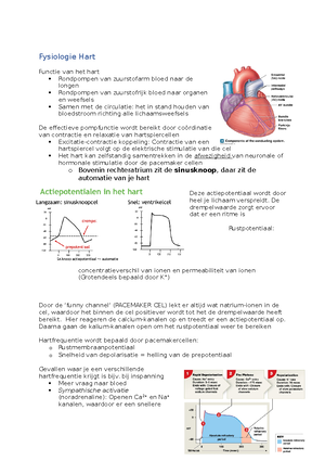
20: The cells of the conducting system distribute electrical impulses through the heart, causing cardiac contractile cells to contract Single cardiac contraction: heartbeat - Pacemaker cells of conducting system control and coordinate heartbeat => contractile cells produce powerful contractions that propel blood - Action potential (electrical impulse) generated and propagated and distributed => triggers contraction of cardiac contractile cells (is lagging) - atria => AV valves => ventricles => semilunar valves Autorhythmicity= cardiac muscle tissue contracts on its own, without neuronal or hormonal stimulations
Pacemaker cells (nodal cells) essential for heart rate - Sinoatrial (SA) node: right atrium - Atrioventricular (AV) node: junction between atria and ventricles => sends on message Conducting cells interconnect the SA and AV node. In atria internodal pathways and in ventricles the AV bundle (bundle of His) and purkinje fibers (larger in diameter) - Excitable membranes of pacemaker cells do not have a stable resting membrane potential = pacemaker potential >> slow inflow of Na+ without compensating outflow of K+
- SA node reaches threshold first: sinus rhythm (SA node brings AV pacemaker cells to threshold faster)
- Ventricular ejection: semilunar valves are pushed open and blood flows into pulmonary and aortic trunks
- stroke volume (SV) of the heart
- Semilunar valves close: end of ventricular systole => ventricular pressures fall rapidly => closes valves
- blood remaining in ventricle when semilunar valve closes= end-systolic volume (ESV)
- Isovolumetric relaxation: all heart valves are closed. Ventricular pressures still higher than atrial, blood cannot flow into ventricles. Ventricular pressures drop rapidly during relaxation
- AV valves open; passive ventricular filling occurs. Atrial pressures force the AV valves open
Listening to the heart: auscultation (with stethoscope) – heart murmur: turbulence of regurgitation (inconsequential)
20: Cardiac output is determined by heart rate and stroke volume Cardiac output (CO): amount of blood pumped by the left ventricle in 1 minute= indication of blood flow through peripheral tissue (ventricular efficiency over time) - Heart rate (HR): number of heart beats per minute – can be adjusted by autonomous nervous system and hormones - Stroke volume (SV): amount of blood pumped out of ventricle during each contraction
CO (mL/ min) = HR (beats/min) * SV (mL/beat) - SV= EDV (end-diastolic volume) – ESV (end-systolic volume) [changes in EDV and ESV can change SV and CO] - Get the largest stroke volume when EDV is as large as possible and ESV is as small as possible
Autonomic activity and circulating hormones make homeostatic adjustments to the heart rate as cardiovascular demands change - Sympathetic and parasympathetic division of autonomic nervous system innervate heart by cardiac plexus - Both ANS division innervate SA and AV nodes and ventricular contractile cells (sympathetic fibers outnumber) Cardiac centers monitor baroreceptors and chemoreceptors – adjust the heart’s activity (dual innervation => resting autonomic tone) - Parasympathetic effects dominate in a healthy, resting individual. If parasympathetic activity increases, the heart rate declines further. ANS changes the ionic permeabilities of cells in the conducting system (especially at SA node) - Cardio acceleratory center controls sympathetic neurons - norepinephrine (binds beta-1 receptors) released by sympathetic neurons opens sodium and calcium ion channels => heart rate increases - has a positive inotropic effect => stimulation increases the metabolism of cardiac contractile cells => ventricles contract more forcefully
- Cardio inhibitory center controls the parasympathetic neurons
- acetylcholine released by parasympathetic neurons opens chemically gated K+ channels in plasma membrane => slowing rates => heart rate declines
- stimulation from the vagus nervus has a negative inotropic effect => produces hyperpolarization and inhibition => force of cardiac contractions is reduced (atria show greatest force)
H19 Blood
19: blood, composed of plasma and formed elements, provides transport, regulation and protective services to the body Blood is a specialized connective tissue and has many vital roles - Transporting dissolved gasses, nutrients, hormones, and metabolic wastes (RBC; oxygen) - Regulating the pH and ion composition of interstitial fluids - Restricting fluid losses at injury sites - Defending against toxins and pathogens (white blood cells; antibodies) - Stabilizing body temperature (blood absorbs the heat from muscles and work)
Whole blood from any source has same basic physical characteristics: 38 degrees, viscous (dissolved proteins), slightly alkaline >> 7 percent of the body weight Whole blood: fluid plasma and formed elements (van be fractioned for analysis) - Plasma proteins - albumins: plasma osmolarity (thyroid hormones) - globulins: immunoglobulins, transport globulins (hormone-binding protein), apolipoprotein - fibrinogen: clotting (removes the clotting proteins, leaving a fluid: serum) Liver synthesizes and releases more than 90 percent of the plasma proteins - Formed elements if blood are made up primarily of red blood cells, white blood cells and platelets - produced in the process of hemopoiesis (45 percent)
19: Red blood cells, formed by erythropoiesis, contain hemoglobin that transport respiratory gasses Red blood cells (RBCs): erythrocytes contain hemoglobin that binds and transports the respiratory gases oxygen and CO2. - Many conditions can affect the hematocrit (blood sample centrifuged) RBCs are a biconcave disc and have a flexible plasma membrane, which pertains to certain functions
- Gives each RCB a large surface-area (oxygen must be absorbed or released quickly)
- Enables RBCs to form stacks that smooth blood flow through narrow blood vessels (=rouleaux)
- Enables RBCs to bend and flex when entering small capillaries (Squeeze)
Mature RBCs are anucleate; have only cytoskeleton => cannot divide => short lifespan - Energy from anaerobic metabolism of glucose
Hemoglobin (Hb) transport oxygen and carbon dioxide - Complex quaternary structure: two alpha and beta chains, all contain heme group which holds an iron ion that interacts with an oxygen => HbO2. - Is reversible; iron-oxygen interaction is weak - Fetal hemoglobin binds oxygen more readily (production can be stimulated in adults with drugs; SCD) Amount of oxygen bound to hemoglobin depends mostly on the oxygen content of the plasma - Bind carbon dioxide: forming carbaminohemoglobin – if hematocrit is low, Hb content of RCBs is reduced (=anemia)
RCB is exposed to severe physical stresses => engulfed => new ones enter bloodstream at comparable size Blood forms primarily in vessels of embryonic yolk sac - Move to liver, spleen, thymus and bone marrow => embryonic cells differentiate into stem cells that divide and produce blood cells - In adults; skeleton enlarges – erythropoiesis takes place in red bone marrow/ myeloid tissue (yellow= fat) - Hemocytoblasts divide and produce 1) myeloid stem cells => red blood cells and 2) lymphoid stem cells => lymphocytes - RCBs first differentiate into proerythroblasts => reticulocyte (after day 4)
Erythropoiesis is stimulated directly by hormone erythropoietin (EPO)= glycoprotein by liver and kidneys - Stimulates cell division rates of erythroblasts; speeds up maturation of RCBs (blood doping) - Low oxygen level in tissues: hypoxia - Red bone marrow needs adequate supplies of amino acids, iron, and vitamins for protein synthesis - need intrinsic factor (produced in stomach) to absorb vitamin B Macrophages of the spleen, liver and red bone marrow => engulf RBCs => remove Hb molecules - Alpha and beta chains eliminated in urine – globular proteins and amino acids are metabolized - Each heme unit is stripped of iron and converted to biliverdin => bilirubin => liver for excretion in bile - bile ducts blocked => bilirubin diffuses => jaundice - Large quantities of free iron are toxic to cells => stored in phagocytic cell => bind to transferrin which can be absorbed by developing red blood cells - Excess transferrins are removed in liver and spleen and stored in ferritin and hemosiderin - iron-deficiency anemia results from a lack of iron in diet or problems with iron absorption
19: Platelets, disc-shaped cell fragments, function in the clotting process Platelets (= thrombocytes) are disc-shaped, continuously replaced and play major role in vascular clotting system, functions: - Releasing chemicals important to the clotting process - Forming a temporary patch in the walls of damaged blood vessels (= platelet plug) - Reducing the size of a break in a vessel wall
Platelet production = thrombocytopoiesis in red bone marrow - Megakaryocytes manufacture structural proteins, enzymes and membranes – loses all of its cytoplasm producing about 4000 platelets
19: The process of blood clotting, or hemostasis, stops blood loss Hemostasis= stopping of bleeding, halts the loss of blood; is in three phases (complex cascade, end is clot retraction)
Vascular phase: local contraction of the vessel is a vascular spasm - Endothelial cells contract and expose the underlying basement membrane to the bloodstream - Endothelial cells begin releasing chemical factors and local hormones (endothelins) - Endothelial plasma membranes become sticky (helps platelets to attach)
Platelet phase: attachment of platelets to sticky endothelial surfaces - Attachment of platelets to exposed surfaces: platelet adhesion => platelet aggregation (positive loop) forms a plug - As platelets arrive at injury site => become activated => releasing compounds - ADP, thromboxane A, clotting factors, PDGF, calcium ions - Several key factors limit growth of platelet plug - prostacyclin, inhibitory compounds (by WBCs), plasma enzymes that breakdown ADP Coagulation phase: blood clotting (until 30 seconds) - Depends on the presence of clotting factors/ procoagulants – are proenzymes => converted to active enzymes => direct clotting response
- Mucosa-associated lymphoid tissue (MALT) > deep to intestine: aggregated lymphoid nodules/ Peyer’s patch - boundaries not distinct => no fibrous capsule - central zone: germinal center; contains dividing lymphocytes Lymphoid organs include the lymph nodes, thymus and spleen
- Lymph nodes: small. Lymphoid organs (1- 25 mm)
- dense connective tissue capsule covers each lymph node
- fibrous partitions: trabeculae
- blood vessels and nerves reach lymph node at hilum
- Afferent lymphatics bring lymph to lymph node (efferent lymphatics leave the lymph node at the hilum)
- Subcapsular space (macrophages and dendritic cells) => cortex (B cells) => paracortex (T cells; lymphocytes enter blood stream) => medulla= core (B-cells and macrophages) >> through network of sinuses
Lymph node purifies lymph before it reaches the veins - Early defense system – antigens into interstitial fluid => into lymph – stimulate macrophages and lymphocytes - Largest lymph nodes located where peripheral lymphatics connect with trunk - Minor infection commonly produces slight enlargement of lymph nodes (increase number of lymphocytes)
Thymus is a primary lymphoid organ (becomes inactive later on): in mediastinum - After puberty it gradually diminishes in size and becomes fatty and fibrous = involution - Capsule cover two thymic lobes - outer cortex densely packed with lymphocytes and a paler, central medulla - epithelial reticular cells maintain blood thymus barrier around blood vessels of cortex => separates developing T cells from the general circulation - Thymosin (hormone) promotes development and maturation of T cells
Adult spleen contains the largest collection of lymphoid tissue in body - (1) Remove abnormal blood cells by phagocytosis (2) storing iron (3) initiating immune responses by B and T cells in response to antigens in circulating blood - Attached to lateral border of the stomach by gastrosplenic ligament - Lymphatic vessels communicate with spleen on visceral surface at the hilum - red pulp (red blood cells and macrophages) and white pulp (white blood cells and lymphocytes) within capsule - Splenic artery => branch into trabecular arteries => branches capillaries surrounded by white pulp => discharge blood into red pulp - Sinusoids lined by macrophages => empty into small veins => collect into trabecular veins Circulatory arrangement gives phagocytes in the spleen the opportunity to identify and engulf damaged or infected cells in circulating blood - Spleen tears easily => internal bleeding and circulatory shock = difficult to repair surgically (sutures tear) - severe ruptured spleen where spleen is removed = splenectomy; but increased risk of bacterial infection
H26 The Urinary System
26: The Organs of the Urinary System function in excreting wastes and regulating body fluids - Paired kidneys have excretory functions, produce urine (contains water, ions, small soluble compounds) Urinary tract: ureters => urinary bladder => urethra
Excretion: removal of metabolic waste products from body fluids and elimination: discharge of these wastes out of body - Regulating blood volume and pressure (volume water lost in urine, release of erythropoietin, renin) - Regulating plasma concentrations of sodium, potassium, chloride and other ions - Helping to stabilize blood pH (concentrations of hydrogen and bicarbonate ions) - Conserving valuable nutrients - Assisting the liver (detoxifying poisons, starvation, deamination of amino acids)
26: Kidneys are highly vascular organs containing functional units called nephrons Kidneys are held in position within abdominal cavity; three concentric layers of connective tissue protect and stabilize each kidney: fibrous capsule (collagen fibers), perinephric fat, renal fascia - Arrangement prevents the jolts and shocks of day-to-day living - Kidneys are served by renal blood vessels, lymphatics, and nerves adjacent to proximal region of ureters Hilum: medial indentation; point of entry for renal artery and nerves, and exit for vein
- Renal cortex (granular): superficial region of the kidney, in contact with the fibrous capsule
- Renal medulla
- renal pyramids (tip of each: papilla – projects in renal sinus)
- renal columns extend into medulla and separate adjacent renal pyramids
- Kidney lobe consists of renal pyramid, the overlying area of renal cortex, and adjacent tissues of renal columns
- Four or five minor calyces merge to form a major calyx – two or three major calyces combine to form renal pelvis => connected to the ureter => drains the kidney
Urine production begins in nephrons – cortical nephrons (renal cortex) and juxtamedullary nephrons (renal medulla)
As it enters the renal sinus, the renal artery provides blood to the five segmental arteries - Numerous afferent arterioles deliver blood to the capillaries supplying individual nephrons - Renal nerves innervate the kidneys and ureters: sympathetic postganglionic fibers
Functional units: nephrons - Renal corpuscle (spherical): contains capillary network that filters blood - glomerular capsule (Bowman’s) envelopes the capillary network: glomerulus - capsule encapsulates the glomerular capillaries and is continuous with initial segment of renal tubule - glomerulus consists of 50 intertwined capillaries – leaves in efferent arteriole - capsular space separates the capsular outer layer and visceral layer
Visceral layer consists of large cells with feet that wrap around dense layer: podocytes - Foot processes, narrow gaps (filtration slits) between adjacent foot processes
Intraglomerular mesangial cells are located among the glomerular capillaries = specialized cells derived from smooth muscle (contraction decreases luminal diameter of capillaries) - Structural support, filtration, phagocytosis - Glomerular capillaries are fenestrated capillaries
Filtration membrane: fenestrated endothelium, basement membrane, foot processes of podocytes - Blood pressure forces water and small dissolved solutes out of the glomerular capillaries through this membrane into the capsular space => proteins-free solution (filtrate)
Renal tubule: long tubular passageway, that begins in corpuscle and ends in collecting system Two convoluted segments - Proximal convoluted tubule: first segment - simple cuboidal epithelium whose apical surfaces have microvilli - reabsorption of critical ions - - filtrate travels along renal tube: tubular fluid; gradually changes in composition (reabsorbed: reclaimed and enters blood) - divided into descending (fluid flows towards renal pelvis) and ascending limb (towards renal cortex) - Distal convoluted tubule: third segment – passes between the afferent and efferent arterioles - opens into the collecting system
Juxtaglomerular complex (structure) helps regulate blood pressure and filtrate formation - Macula densa: group closely packed epithelial cells in distal tubular epithelium - Juxtaglomerular cells= baroreceptors: modified smooth muscle cells in wall of afferent arteriole that secrete renin - Extraglomerular mesangial cells (located in triangular space between afferent and efferent glomerular arterioles): provide feedback control between macula dense and juxtaglomerular cells
Collecting system begins in cortex and descends into medulla – and transports tubular fluid from nephrons to renal pelvis Adjusts fluid’s composition and determines final osmotic concentration and volume of urine - Several collecting ducts converge into larger papillary duct - intercalated cells: cuboidal cells with microvilli - principal cells: cuboidal cells that lack microvilli and reabsorb water and secrete potassium ions
Cortical nephrons (85%) - Nephron loop is short and efferent arteriole delivers blood to a network of peritubular capillaries Juxtamedullary nephrons (15%) - Long nephron loops that extend deep into the renal pyramids of the medulla – efferent arterioles connect to vasa recta
26: different segments of the nephron from urine by filtration, reabsorption and secretion Urine production is to maintain homeostasis by regulating the volume and composition of the blood (metabolic wastes) - Urea (most abundant metabolic waste) - Skeletal muscle tissue generates creatinine (role in muscle contraction) - Uric acid: waste formed during recycling of nitrogenous bases from RNA molecules Wastes are delivered in bloodstream => their removal involves loss of water If kidneys were unable to concentrate the filtrate produced by filtration, fluid losses would lead to fatal dehydration in hours
Processes of urine formation 1) Filtration—blood pressure forces water and solutes across the walls of glomerular capillaries and into capsular space >> based solely on particle size 2) Reabsorption: removal of water and solutes from filtrate =>into peritubular fluid; takes place after filtrate has left renal corpuscle >> selective process (involves simple diffusion or carrier proteins)
Summary Anatomy and Fysiology
Vak: Anatomie en fysiologie (AB_1176)
Universiteit: Vrije Universiteit Amsterdam

- Ontdek meer van: